

Translate this page into:
Dysmenorrhea membranacea: A case report and review of the literature
This is an open access journal, and articles are distributed under the terms of the Creative Commons Attribution-NonCommercial-ShareAlike 4.0 License, which allows others to remix, tweak, and build upon the work non-commercially, as long as appropriate credit is given and the new creations are licensed under the identical terms.
This article was originally published by Wolters Kluwer - Medknow and was migrated to Scientific Scholar after the change of Publisher.
Abstract
Dysmenorrhoea membranacea is a rare condition in which there is spontaneous expulsion of the fragments of endometrium in a cylindrical piece retaining the shape of the uterus. Here we present a case of dysmenorrhoea membranacea in 53 year old female patient who was on hormonal therapy for her irregular menstrual history, presented with the history of menorrhagia and passage of clots since one month.. Patient was subjected to D&C, during PV examination there was an expulsion of this endometrial cast. On histopathology it was diagnosed as dysmenorrhoea membranacea. It is an uncommon diagnosis, predominantly occurring in second and third decade of life. We also reviewed the literature associated with this lesion.
Keywords
Dysmenorrhea membranacea
endometrial hyperplasia
menorrhagia

INTRODUCTION
Dysmenorrhea membranacea is a clinicopathological term first described by G.B. Morgagni in 18 th century. Clinically, there is shedding of the endometrium in a shape resembling the uterus.[1] It can cause intense cramping pain because the intact cast passes through an undilated cervix.[2] Histopathologically, decidualization of endometrial stroma and atrophy of endometrial glands is seen. Exact pathophysiology is not known, but high progesterone levels have been thought as one of the etiological factors. Only 16 cases had been reported till 2009.[1]
CASE REPORT
A 53-year-old female, para five, living four presented with menorrhagia and passage of clots since 1 month. Patient was on hormonal therapy. Ultrasonography showed thickened endometrium and suggestive of hyperplasia. Patient was subjected to hysteroscopic Dilation and Curettage, while pelvic examination there was an expulsion of mass which was sent for histopathological examination.
HISTOPATHOLOGICAL FINDINGS
Grossly specimen consisted of a grey brown degenerated tissue mass measuring 22 cm × 8 cm × 2 cm. External surface was gray-brown, irregular with small cystic areas in between. Cut surface was grey white and micro cysts were seen [Figure 1].
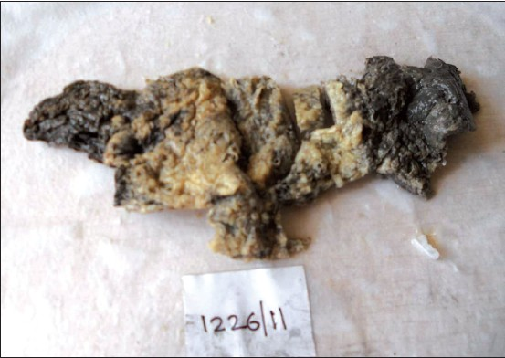
- Gross photo showing grey brown tissue with shape of uterus
On microscopy, cystically dilated endometrial glands, large areas of decidualized endometrial stroma, hemorrhage, and acute inflammatory cell infiltrate were seen [Figures 2-3]. Based on these findings and clinical history, diagnosis of dysmenorrhea membranacea was done.

- Decidualized stroma with cystically dilated glands (H and E, ×5)

- Decidualized stroma with blood vessels (H and E, ×10)
DISCUSSION
Dysmenorrhea membranacea is a clinical term involving spontaneous shedding of the endometrium in one or several membranous pieces retaining the shape of the uterine cavity. It is considered as a variety of irregular shedding of the endometrium.[3] Though rare, it is one of the causes of secondary dysmenorrhea.[4] Clinical features include cramping pain during expulsion of the cast.
Many hypotheses have been proposed for the etiology. Greenblatt et al. suggested the cause being raised production of estrogen and progesterone with partial dissolution of thickened endometrium. Another theory suggested a normal but intense development of spiral arteries with excess vasodilatation and then intense vasoconstriction with shedding of an overdeveloped endometrium. Dallenback-Hellweg stated the condition being of hyperprogesteronism which can be caused exogenously or endogenously like excess production of hypophyseal gonadotropins or hyperfunctioning corpus luteum. In this condition, dysmenorrhea occurs due to the failure of the tissue to undergo dissolution while the spontaneous detachment of intact cast can be explained by focal release of relaxin at the demarcation line and comparable with detachment process after delivery.[3]
This condition is seen mainly in young women between 20 and 40 years. Dysmenorrhea may or may not be present. Most of the cases had a history of hormonal therapy, but cases without hormonal therapy were also reported. In this case, age of the patient is 53 years which is rare. The patient had dysmenorrhea and history of hormonal intake.
Usually, the diagnosis of this condition is established on histopathological examination of expulsed tissue or after endometrial curettage.[1] On routine Hematoxylin and Eosin stained sections we see decidualized endometrium with regressive changes or focal hemorrhagic necrosis and infiltrated by neutrophils. The decidual cells may have a spindly shape when regression is advanced. The endometrial glands are lined by cuboidal epithelium. In this case, the histological features match with the routine findings.
This condition has a well-known association with ectopic pregnancy. Ectopic pregnancy with cast can be mistakenly diagnosed as intrauterine pregnancy on ultrasonography. The cast expulsion can be wrongfully thought as miscarriage clinically. It can also be seen in nonpregnant women as an adverse effect of human menopausal gonadotropin, human chorionic gonadotrophin, and progestogens.[5] In this case, there was no ectopic pregnancy. However, there was the intake of progestogens.
We conclude that in nonpregnant women on hormonal therapy or oral contraceptives with complaints of severe cramping abdominal pain and bleeding with passage of tissue, dysmenorrhea membranacea, though rare, should be thought as a diagnosis. Moreover, this condition can be wrongfully diagnosed as intrauterine pregnancy on ultrasonography in undiagnosed ectopic pregnancies. In such cases, follow-up with serial beta hCG levels should be done.
Source of Support:
Nil
Conflict of Interest:
None declared.
REFERENCES
- Chronic fatigue syndrome: A hormonal origin? A rare case of dysmenorrhea membranacea. Arch Gynecol Obstet. 2009;279:717-20.
- [Google Scholar]
- Functional disturbances of the endometrium. Haines and Taylor Obstetrical and Gynaecological Pathology. (3 rd). :334-5.
- [Google Scholar]


